3D Pre-Operative Visualisation Tool
AI-powered 3D visualisation that helps surgeons plan complex procedures faster, safer, and with greater precision.
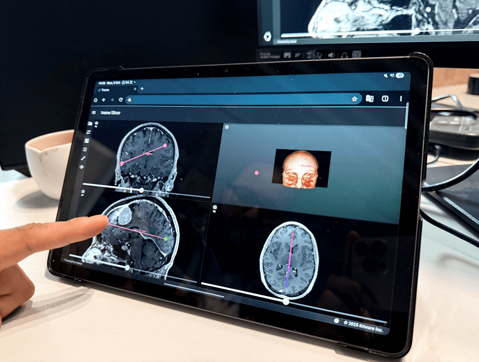
OneBonsai developed a proof-of-concept 3D visualization tool to support neurosurgeons in planning brain tumor surgeries. Using AI-powered segmentation of CT scans, the system generates an interactive 3D model of the tumor and surrounding anatomy, which can be accessed securely on a tablet. This intuitive approach simplifies preoperative planning, reduces preparation time, and enhances spatial understanding of tumor morphology, giving surgeons clearer insights to make safer and more precise decisions.
Simplifying 3D Tumor Visualization for Surgeons
Challenge
Intuitive 3D Tumor Visualization Tool
Solution
Collaboration with Medical Experts and Advanced Tech
Approach
A Promising Proof of Concept
Outcome
Challenge
Neurosurgeons planning brain tumor operations traditionally rely on 2D CT scan slices to understand the size, shape, and position of tumors relative to surrounding brain structures. This approach requires significant mental effort to reconstruct a three-dimensional picture from flat images, which can be time-consuming and prone to misinterpretation. For complex cases, the limitations of 2D imaging can make it difficult to fully appreciate tumor morphology and plan optimal surgical approaches. There was a clear need for an intuitive 3D visualization tool that could simplify preoperative planning and improve surgical precision.

“Relying only on 2D CT scans made it difficult for surgeons to clearly understand tumor size and position before operating.”
Solution
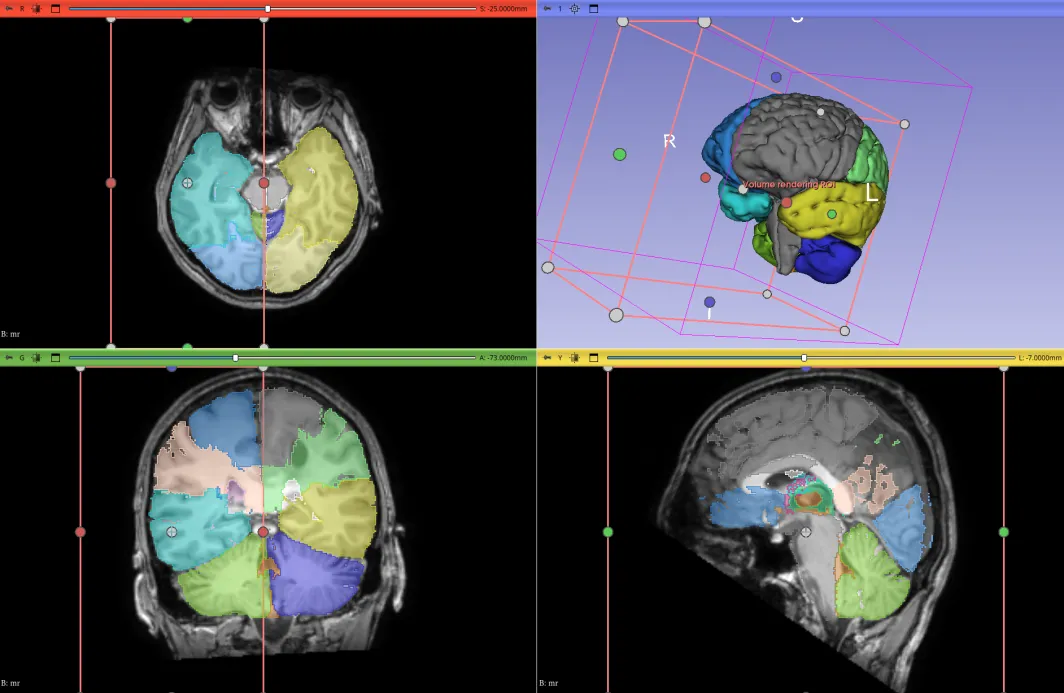
OneBonsai developed a proof-of-concept 3D visualization tool that uses AI-powered segmentation to automatically process CT scan data and generate interactive 3D models of brain tumors and surrounding anatomy. The resulting model can be viewed and manipulated on a standard tablet device. Surgeons can rotate, zoom, and explore the 3D model from any angle, gaining a comprehensive spatial understanding of the tumor's relationship to critical brain structures. The tool is designed to be intuitive and accessible, requiring no specialized hardware beyond a tablet with secure access.
Approach
The development of the 3D visualization tool involved tight collaboration between OneBonsai's technical team and neurosurgical experts. Medical professionals provided guidance on the most critical visualization requirements and validated the accuracy of the AI-generated segmentation results. The AI segmentation pipeline was trained on medical imaging data to accurately identify and delineate tumor boundaries. The interactive 3D viewer was optimized for tablet performance, ensuring smooth, responsive interaction with complex anatomical models while maintaining data security and patient privacy.
“Reviewing a tumor in 3D can now be done quickly and easily, further improving patient care.”
Outcome
The 3D Pre-Operative Visualisation Tool demonstrated significant potential for improving neurosurgical planning. Surgeons who tested the prototype reported faster preparation times, better spatial understanding of tumor anatomy, and increased confidence in their surgical approach. The proof of concept validated the feasibility of AI-powered 3D medical visualization as a practical clinical tool. The project established a strong foundation for further development and clinical validation, with the potential to transform preoperative planning across multiple surgical specialties.
3D Pre-Operative Visualisation Tool

3D Pre-Operative Visualisation Tool
“The tool saves time in preparation and gives our surgeons a new level of precision.”
Frequently Asked Questions
More Projects